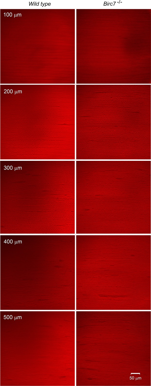
Figure 12.

Multiphoton microscopic analysis of cellular organization in lenses from wild-type or Birc7-null animals. Crosses were established with mT/mG mice (a line in which the fiber cell membranes are endogenously labeled with TdTomato) to allow the morphology of individual cells located at various depths below the lens surface to be visualized. Individual optical sections are shown. Note that the border of the OFZ is located approximately 200 μm below the lens equatorial surface. There was no obvious difference in cell morphology or packing arrangement on either side of the OFZ border.
