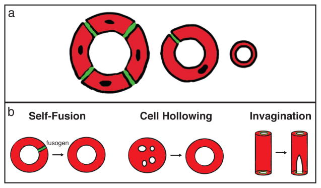
Figure 1. Formation of seamless tubes.

Tube architectures defined by the presence and number of cell junctions are schematized in cross-section (a). Multicellular tubes (left) are comprised of 2 or more cells surrounding a lumenal space and connected by intercellular junctions (green). Auto-cellular tubes (middle) are single cells wrapped around a lumenal space and sealed into a tube by self-junctions (green). Seamless tubes (right) have an intracellular lumen bounded by an internal apical membrane. In (b), three mechanisms of seamless tube formation are illustrated: (left) in self-fusion, the junctional seam of an autocellular tube is removed in a fusogen-dependent step; (middle) cell hollowing, in which large membrane-bound bodies (“vacuoles”) coalesce and fuse in the middle of the cell; (right) invagination, where a small apical domain demarcated by inter-cellular junctions is extended internally
