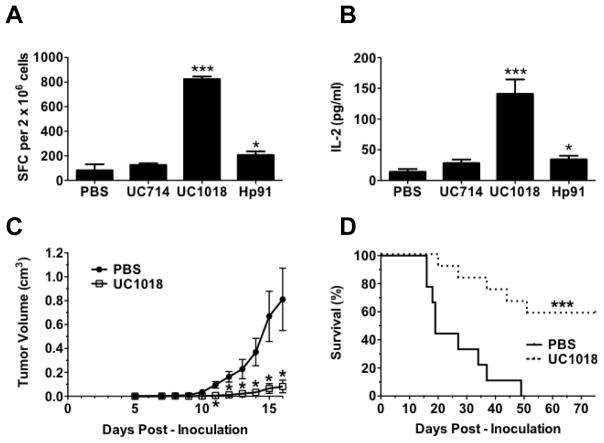
Figure 5. The short alpha helical peptide UC1018 induces protective immune responses in vivo.
(A, B) Mice were co-immunized with OVA-I (SIINFEKL) peptide with PBS (negative) or equimolar doses of Hp91 (250μg), UC714 (129μg), or UC1018 (142μg). Freshly isolated splenocytes from the immunized mice were cultured in the presence of SIINFEKL peptide (2.5μg/ml) in an (A) IFN-γ ELISpot assay, wherein the number of IFN-γ-secreting cells, or spot-forming cells (SFC), was determined 18 h later, or (B) culture supernatants were collected and analyzed for IL-2 secretion by ELISA. The data are shown as mean (±SEM) for at least 5 mice/group. *p < 0.05, or *** p < 0.001 compared to PBS; Student’s t-test. (C, D) Mice were immunized s.c. with apoptotic mitomycin-C treated B16 cells co-injected with PBS (negative) or UC1018 peptide adjuvant (142μg) and boosted twice. One week post boost, mice were inoculated s.c. on the flank with 5×10⁵ live B16 cells. (C) Tumor dimensions were measured over time. The data shown is mean (±SEM) for at least 14 mice/group. *p < 0.01 compared to PBS; Student’s t-test at each time point. (D) Depicted is the percentage of surviving mice. Mice were euthanized when tumor volume reached 1.5 cm3. Tumor survival curves were generated, wherein the day of euthanasia was considered as death. ***p < 0.001 compared to PBS; Log rank test.

