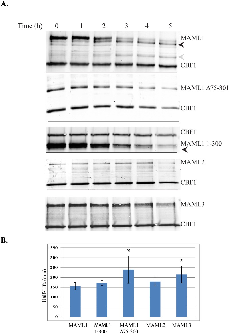
Fig 3. Half-life studies to MAML1-3 family members.
(3A) MAML1, MAML1Δ75–301, MAML1-1-300, MAML2 or MAML3 were expressed with myc-tagged CBF1 in HeLa cells. Cells were treated with 150 μg/ml cycloheximide and cell extracts collected every hour for 5 hours. Western blots were performed to the myc-tag for the various proteins or to endogenously expressed GAPDH. Results shown are from representative blots form three different experiments. Black and gray arrowheads indicate degradation products that could be detected with time with the MAML1 and MAML1-300 constructs. (3B) ImageJ software was used to quantitate expression levels normalized to either CBF1 or GAPDH. The data is presented as the mean ± SD in bar graphs. A * indicates significantly different compared to MAML1 (p<0.05).

