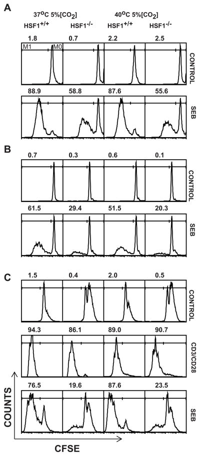
FIGURE 4.
HSF1−/− T cells show compromised proliferation in vitro in response to SEB simulation at both physiologic and febrile temperatures. CFSE labeled spleen cells (A) or Lymph node cells (B) from HSF1−/− or HSF1+/+ mice were cultured in media alone or with 20μg/ml SEB in 24 well plates for 3 days at 37°C or 40°C and 5% CO2. (C) CFSE labeled spleen cells from HSF1−/− or HSF1+/+ mice were activated at indicated temperatures, either alone (Control), or with anti-CD3 and anti-CD28 Abs or SEB (20μg/ml). Following activation, cultures were harvested, washed, labeled with anti-Vβ8 and anti-CD3 and CFSE profiles on live gated Vβ8+CD3+ T cells were analyzed using FACScan® flow cytometer. Numbers above each histogram represent the percent of total gated live cells (average of three replicates) which have undergone cell division (M1 gate, left line). Histograms are representative of two (C) or three (A, B) independent experiments with 3 replicates in each treatment. In each experiment cells were obtained from 2 mice in each strain.

