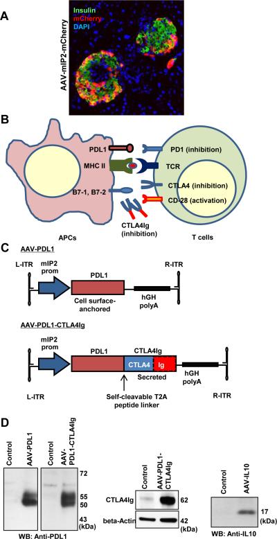
Figure 1. Generation of a β-cell-targeted AAV vector expressing an artificial PDL1-CTLA4Ig.
A. Pancreas sections were observed for mCherry expression. β-cell-specific mCherry expression (red) was observed in insulin-positive β-cells (green), but not in acinar cells. Nuclei were counterstained by DAPI (blue). B. Schematic representation of immuno modulatory molecules, including CTLA4Ig protein. C. Schematic representation of AAV vectors. D. Western blotting analysis verified expression of PDL1, CTLA4Ig and IL10 by specific antibodies.

