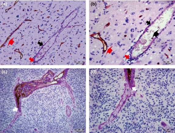
Fig 1.

Relationship between vasculogenic mimicry (VM) and clinicopathological data in glioma tissues. (a, b) Endothelial cells were detected by anti-CD34 immunohistochemistry staining, resulting in a brown (or some black-brown) product. Periodic acid–Schiff (PAS)-positive substances formed a basement membrane-like structure that is highlighted pink or pink-purple (thick black arrows). Representative VM channels are lined with tumor cells (thick red arrows) and rich in PAS+ substances, whereas the luminal surfaces of PAS+ channels (thick black arrows) were negative for CD34 reaction. In the hollows, red blood cells (bold black arrow) can be observed. Typical blood vessels (bold red arrows) showed a positive reaction for CD34 on their luminal surface and PAS+ reaction in their wall. (b) is a magnified image of (a). (a) Scale bar = 100 μm (×200). (b) Scale bar = 50 μm (×400). (c, d) Some VM structures were interlinked with CD34+ endothelial cell-lined blood vessels (white arrows). (d) is a magnified figure of (c). (c) Scale bar = 100 μm (×200). (d) Scale bar = 50 μm (×400). All sections were stained with CD34 and PAS.
