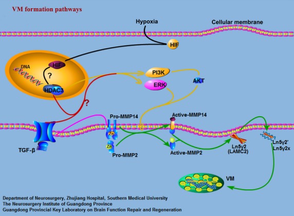
Fig 7.

Signaling pathways implicated in the formation of vasculogenic mimicry (VM) networks. Previous observations have suggested a classical model of the signaling cascade implicated in VM. Green arrows indicate the classical signaling pathways implicated in VM in gliomas. In this study, we showed that histone deacetylase 3 (HDAC3) can regulate VM through the phosphoinositide 3-kinase (PI3K)/ERK–MMPs–laminin5γ2 (Ln5γ2) signaling pathways in gliomas. HDAC3 can first regulate the activation of ERK1/2 and PI3K directly or indirectly (yellow arrows). Active PI3K then regulates directly or indirectly the transition of pro-MMP14 into active MMP-14, which subsequently activates pro-MMP2 (green arrows). Both MMP-14 and MMP-2 can promote the cleavage of Ln5γ2 into the pro-migratory fragments 5γ2′ and 5γ2x (green arrows). These fragments result in the formation of VM networks. We have previously shown that transforming growth factor-β (TGF-β) can regulate VM by MMP-14 (pink arrows). However, HDAC3 is need for TGF-β to regulate the activation of the ERK and PI3K signaling pathways. Hence, we may further investigate whether HDAC3 pathways are involved in the process by which TGF-β (red arrows) regulates VM. Other studies reported that hypoxia-inducible factor-1α (HIF-1α) has been shown to induce VM and, interestingly, HDAC3 has been described as a HIF-1α regulated gene. Also, hypoxia enhances HDAC function, such that HDACs are closely involved in angiogenesis. Hence, it would be of interest to investigate whether the HDAC3 pathway is also involved in processes where hypoxia regulates VM (black arrows). AKT, protein kinase B; LAMC2, laminin, γ2.
