
Fig 2.
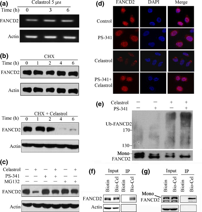
Celastrol triggers FANCD2 degradation through the ubiquitin–proteasome pathway. (a) RT-PCR assays for detecting FANCD2 RNA level in A549 cells upon celastrol. (b) Effects of cycloheximide (CHX) (50 μg/mL) alone or in combination with celastrol (5 μM) on FANCD2 expression, evaluated by Western blotting in A549 cells. (c) A549 cells were pre-incubated with MG132 (10 μM) or PS341 (100 nM) for 1 h, followed by celastrol (5 μM) treatment for 6 h. Cell lysates were subjected to Western blotting using anti-FANCD2 antibody. (d) A549 cells were pretreated with PS341 (100 nM) for 1 h, followed by celastrol (5 μM) incubation for 6 h. The cells were then analyzed by immunofluorescence assay labeling with anti-FANCD2 antibody and DAPI. (e) A549 cells were pretreated with or without PS341 (100 nM) for 1 h, followed by celastrol incubation for 2 h. Cell lysates were subjected to immunoprecipitation with FANCD2 antibody, followed by Western blotting using antibodies against FANCD2 and ubiquitin. (f, g) A549 cells were treated with Biotin or Bio-Cel at 50 μM for 4 h, lysed, and the cell lysates were subjected to immunoprecipitation using streptavidin agarose and Western blotting using indicated antibodies. Mono, monoubiquitinated FANCD2.