Fig 3.
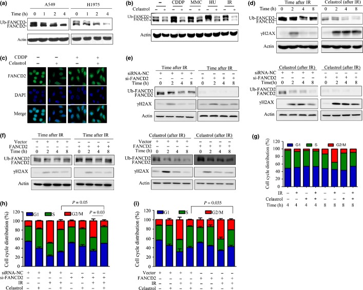
Celastrol downregulates monoubiquitinated FANCD2 and attenuates DNA-damaging agent-induced FA pathway activation. (a) A549 and H1975 cells were treated with celastrol at 5 μM for indicated time points, lysed, and Western blotting was performed using anti-FANCD2 antibody. (b) FANCD2 monoubiquitination induced by different DNA-damaging agents was inhibited by celastrol. A549 cells were pre-incubated with cisplatin (CDDP; 20 μM/L), mitomycin C (MMC) (0.4 μg/mL) or hydroxyurea (HU; 1 mmol/L) for 24 h or subjected to ionizing radiation (IR) (15 Gy), then incubated with celastrol (5 μM) for additional 4 h. The cells were lysed and Western blot was performed. (c) A549 cells were pretreated with cisplatin (20 μM/L) for 24 h, followed by celastrol (5 μM) incubation for additional 4 h. The cells were subjected to immunofluorescence assay by labeling with anti-FANCD2 antibody and DAPI. (d) A549 cells were irradiated by X-ray (15 Gy), then incubated immediately with 5 μM celastrol at indicated time points; whole-cells extracts were used for detection of FANCD2 and γ-H2AX. (e) A549 cells were transfected with siRNA against negative control (siRNA-NC) or FANCD2 (siFANCD2), irradiated by X-ray (15 Gy), then incubated immediately with 5 μM celastrol at indicated time points. The whole-cells extracts were used for detection of FANCD2 and γ-H2AX. (f) A549 cells were transfected with pDONR201-vector or pDONR201-FANCD2, irradiated by X-ray (15 Gy), then incubated immediately with 5 μM celastrol at indicated time points, and the whole-cells extracts were used for detection of FANCD2 and γ-H2AX. (g) A549 cells were irradiated by X-ray (15 Gy), incubated immediately with 5 μM celastrol for an additional 4 or 8 h. Cell cycle distribution was then determined. (h) A549 cells were transfected with siRNA-NC or si-FANCD2, irradiated by X-ray (15 Gy), then incubated immediately with 5 μM celastrol for additional 8 h. Cell cycle distribution was then determined. (i) A549 cells were transfected with pDONR201-vector or pDONR201-FANCD2, irradiated by X-ray (15 Gy), and then incubated immediately with 5 μM celastrol for an additional 8 h. Cell cycle distribution was determined.

