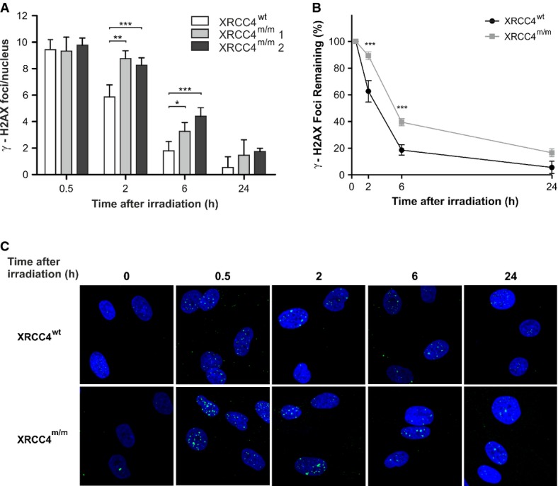
Figure 2.

- γ-H2AX nuclear foci in XRCC4wt and mutant fibroblasts (XRCC4m/m1 and XRCC4m/m2) were determined at different times after irradiation with 0.5 Gy of γ-rays. Values, subtracted of their non-irradiated control (1.3 foci/nucleus in XRCC4wt cells and 1.4 and 0.9 foci/nucleus in XRCC4m/m1 and XRCC4m/m2 cells, respectively), are the mean ± SD of three independent experiments.
- Percentages of γ-H2AX foci in XRCC4wt and XRCC4m/m fibroblasts (pooled values) remaining at the indicated time-points.
- Immunofluorescence micrographs of γ-H2AX foci in XRCC4wt and XRCC4m/m fibroblasts.
Data information: ***P < 0.001; **P < 0.01; *P < 0.05 (XRCC4m/m vs. XRCC4wt cells), two-way ANOVA, Bonferroni post hoc test.
