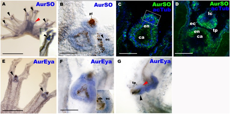
Fig 3. AurSO and AurEya mRNAs are co-expressed throughout the rhopalial ectoderm including the developing photosensory and mechanosensory tissues.
Aurelia sp.1 free-swimming ephyrae were labeled with antisense riboprobes against AurSO (A-D), and AurEya (E-G). In C and D, the rhopalia were also labeled with an antibody against acetylated ∂-tubulin (acTub). A is a lateral view of an ephyra with the mouth/manubrium facing upwards. An inset in A, and B-F are oral views with the rhopalial distal ends pointed upwards. G is a lateral view with the rhopalial distal ends pointed to the left. Note that AurSO and AurEya transcripts strongly localize to rhopalia (black arrowheads in A and E, respectively). Lower levels of manubrial expression were also detected for AurSO (red arrowhead in A), consistent with the result of the previous study [16], and for AurEya (S5 Fig). B: a medial optical section of a rhopalium. An inset shows AurSO expression in both ectoderm (ec) and endoderm (en) separated by mesoglea (arrowhead) in the proximal-lateral region of the rhopalium. C, D: confocal section of rhopalia at the plane of the developing photosensory domain (boxed) (C), and at the plane of the mechanosensory touch plate (tp; D). F: a medial optical section of a rhopalium. An inset shows AurEya expression in ectoderm (ec) but not in endoderm (en), in the proximal-lateral region of the rhopalium. G: a sagittal optical section of a rhopalium, showing AurEya expression in the developing photosensory domain (black arrowhead) and the mechanosensory touch plate (red arrowhead). Abbreviations: pi pigmented endoderm of the cup ocellus; lc lithocyst; ec ectoderm; en endoderm; ca rhopalar canal. Scale bars: 1 mm (A), 500 μm (E), 50 μm (B-D, F, G).

