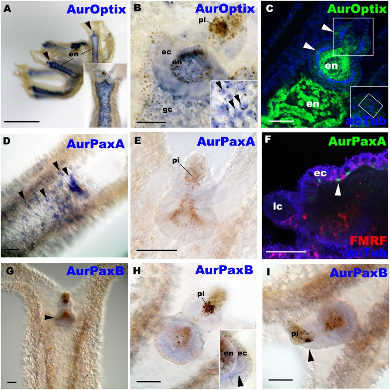
Fig 4. AurOptix, AurPaxA and AurPaxB mRNAs are differentially expressed outside the domain of eye development.
Aurelia sp.1 free-swimming ephyrae were labeled with antisense riboprobes against AurOptix (A-C), AurPaxA (D-F), and AurPaxB (G-I). A is a lateral view of an ephyra with the mouth/manubrium facing upwards. Black arrowheads show rhopalia. Note strong localization of AurOptix transcripts in the endoderm of the arms (en). In C and F, the rhopalia were also labeled with antibodies against acetylated ∂-tubulin (acTub) and FMRFamide-like neuropeptides (FMRF; in F). B, C, E, G and H, and an inset in A, are oral views with the rhopalial distal ends pointed upwards. D is an aboral view with the rhopalial distal end pointed upwards. F and I are lateral views with the rhopalial distal ends pointed to the left. B: a medial optical section of a rhopalium, showing endodermal AurOptix expression. An inset shows AurOptix-expressing endodermal pigment cells (arrowheads) in the gastrovascular canal (gc). C: a medial confocal section of a rhopalium showing AurOptix-expressing cells morphologically identifiable as a sensory cell (upper arrowhead) and ganglion cells (lower arrowhead) (cf. [18]). An inset is the section at the plane of the developing photosensory domain (boxed), showing little AurOptix expression. D: an optical section through the exumbrellar (aboral) ectoderm, showing strong AurPaxA expression in individual cells (arrowheads). E: a medial optical section of a rhopalium, showing no detectable levels of AurPaxA expression. F: a sagital confocal section of a rhopalium and the overlying exumbrellar ectoderm showing that AurPaxA-expressing cells are located at the base of the aboral ectoderm (arrowhead) in close association with the FMRFamide-immunoreactive neuronal network (FMRF). G: an oral view of a rhopalar arm. AurPaxB transcript localization in the basal region of a rhopalium is detectable at low levels (arrowhead). H: a medial optical section of a rhopalium, showing AurPaxB expression in the ectoderm (ec) of the proximal region of the rhopalium (arrowhead in an inset). I: a sagittal optical section through the pigment-cup ocellus of a rhopalium, showing little AurPaxB expression in the developing photosensory domain (arrowhead). Abbreviations: lc lithocyst. Scale bars: 1 mm (A) 50 μm (B-I).

