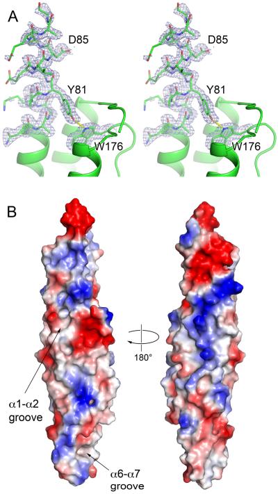
Fig. 4.
The YxxxD/E secretion motif and surface features of EspB.
A. Stereo views of YxxxD/E motif of EspB7-278 (PDB: 4XXX). Residues 76–89 and W176 are shown in stick representation with sA-weighted 2FO-FC electron density map contoured at 1σ. The rest of protein is shown in cartoon representation.
B. Representation of surface charge distribution of EspB7-278 generated using PyMol. The putative lipid-binding sites of EspB are indicated.

