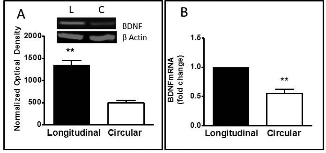
Figure 2.
Expression of BDNF protein and mRNA in smooth muscle cells of rabbit intestine. (A) Western blot of BDNF protein in homogenates of longitudinal and circular muscle cells. Insert illustrates a typical Western blot of BDNF and β actin. Bar graph illustrates optical density of BDNF blots normalized to β actin. Values are Mean ± SEM; n=8; **= p<0.01 for difference between circular and longitudinal muscle. (B) Quantative rt-PCR of BDNF mRNA in circular and longitudinal muscle cells. Relative expression was calculated by the ΔΔCT method as described in Methods. Values are Mean± SEM; n=5; **= p<0.01 for difference between circular and longitudinal muscle.

