. 2015 Jul 21;29(4):233–240. doi: 10.3341/kjo.2015.29.4.233

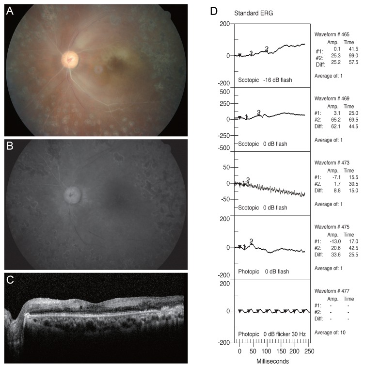
Fig. 3. Images of patient 3, who presented with severe visual decline resulting in no light perception after vitrectomy with retrobulbar anesthesia for proliferative diabetic retinopathy and vitreous hemorrhage. All images were taken 7 days after surgery. (A) A cherry-red spot and severe arterial narrowing and sclerosis are observed in the fundus photograph. (B) Early-phase (2 : 11) fluorescein angiogram showing poor arterial filling. (C) Increased reflectivity and thickness of the inner retina are apparent in spectral domain optical coherence tomography. (D) Standard electroretinogram (ERG) in the left eye showing decreases in both scotopic and photopic wave amplitude.

Fig. 3