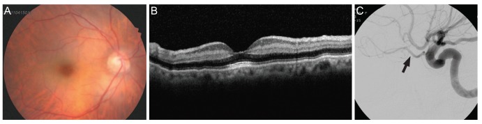
Fig. 5. Images of patient 5, who underwent cataract surgery (phacoemulsification and posterior chamber intraocular lens implant) under retrobulbar anesthesia and presented with visual decline 1 day after surgery. (A) Fundus photograph 1 day after surgery, showing a typical cherry red spot in the right eye. (B) Increased retinal reflectivity and internal retinal thickness are apparent in spectral domain optical coherence tomography. (C) Transfemoral cerebral angiogram performed 1 day after surgery, showing no definitive occlusion in the ophthalmic artery (arrow) or cerebral arteries.