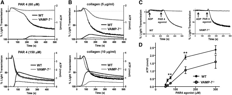
Figure 2.
Dense granule release and platelet aggregation are impaired in VAMP-7−/− platelets. (A) Platelet aggregation (top tracings) and dense granule release (bottom tracings) were monitored in wild-type (black tracings) or VAMP-7−/− platelets (gray tracings) in response to either low concentrations (60 μM) or intermediate concentrations (150 μM) of AYPGKF. (B) Platelet aggregation (top tracings) and dense granule release (bottom tracings) were monitored in wild-type (black tracings) or VAMP-7−/− platelets (gray tracings) in response to either 5 μg/mL or 10 μg/mL collagen. (C) Wild-type (left) and VAMP-7−/− platelets (right) were incubated with a subthreshold concentration of ADP (2 μM) prior to stimulation with AYPGKF (60 μM). Exposure to ADP reverses the aggregation defect in VAMP-7−/− platelets (black tracings). Platelets exposed to ADP alone failed to aggregate (gray tracings). (D) Dense granule release was monitored in wild-type (●) and VAMP-7−/− (○) platelets in response to the indicated concentrations of AYPGKF (**P < .01; n = 3-5). ATP, adenosine triphosphate.

