
Fig. 1.
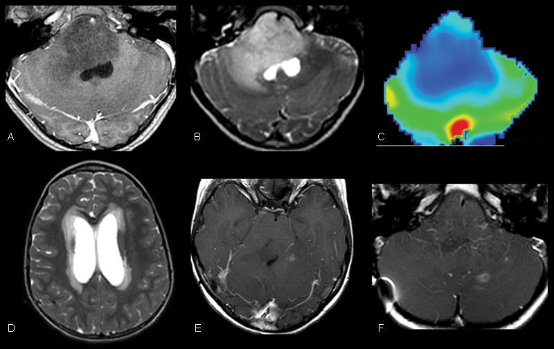
Initial magnetic resonance imaging of our patient demonstrated a grade 3 anaplastic astrocytoma centered within the pons. (A, B) T1 postcontrast and T2-weighted images demonstrate an expansile, nonenhancing, T2 hyperintense mass centered within the pons. (C) Dynamic T2* susceptibility weighted contrast-enhanced cerebral blood volume map demonstrates no evidence of increased cerebral blood volume within the pons consistent with the provided diagnosis. (D) Axial T2-weighted image through the level of the lateral ventricles demonstrates hydrocephalus with transependymal flow of cerebrospinal fluid resulting from the mass effect of the lesion in the midbrain. (E, F) Follow-up surveillance imaging after medial and radiation therapy demonstrates rim-enhancing foci within the supra- and infratentorial brain that suggested the progression of disease.