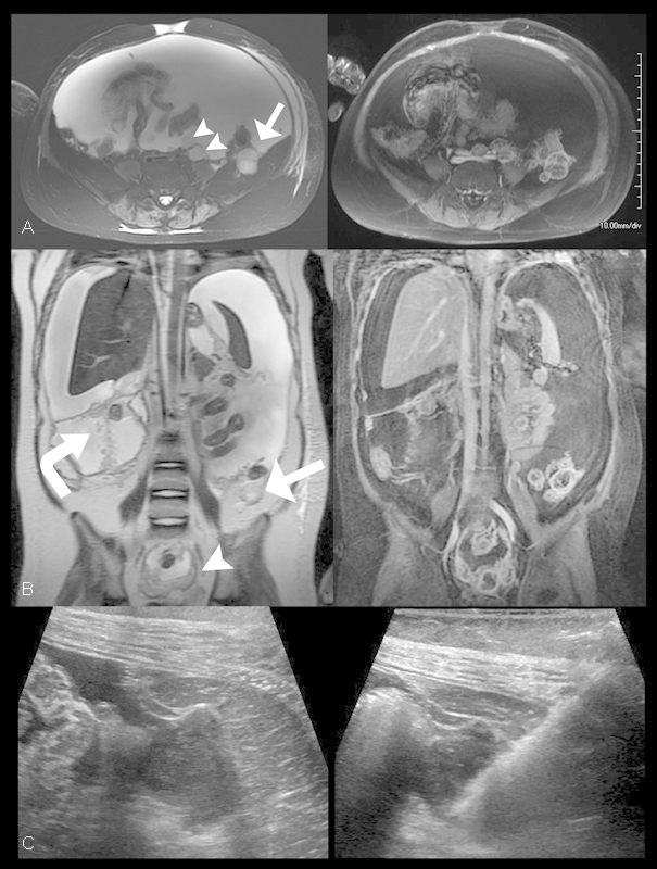
Fig. 2.

Glioblastoma peritoneal carcinomatosis. (A, B) Axial and coronal T2 (left) and T1-weighted fat-saturated postcontrast (right) images through the abdomen and pelvis demonstrates multiple foci of T2 hyperintense enhancing peritoneal tissue nodules (arrowheads) the largest of which was located posterior to the left colon about the Iliacus muscle (arrow) with associated large-volume ascites. Additionally, the VP shunt catheter course was noted to be studded with nodular enhancing foci (curved arrow). (C) Axial pre (left) and post (right) biopsy sonographic images through the left lower quadrant of the abdomen demonstrates the previously observed left paracolic mass anterior to the left Iliacus muscle characterized by predominantly hypoechoic features. The hyperechoic linear lesion corresponds to the core biopsy needle within the mass during ultrasound-guided tissue sampling.
