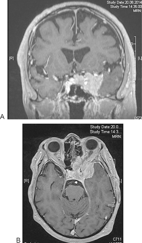
Fig. 4.

Preoperative (A) coronal and (B) axial magnetic resonance imaging (with contrast) of a patient with left-sided medial sphenoid wing meningioma with extension to the orbital, sellar, and ethmoidal region as well as involvement of the cavernous sinus.
