Fig. 4.

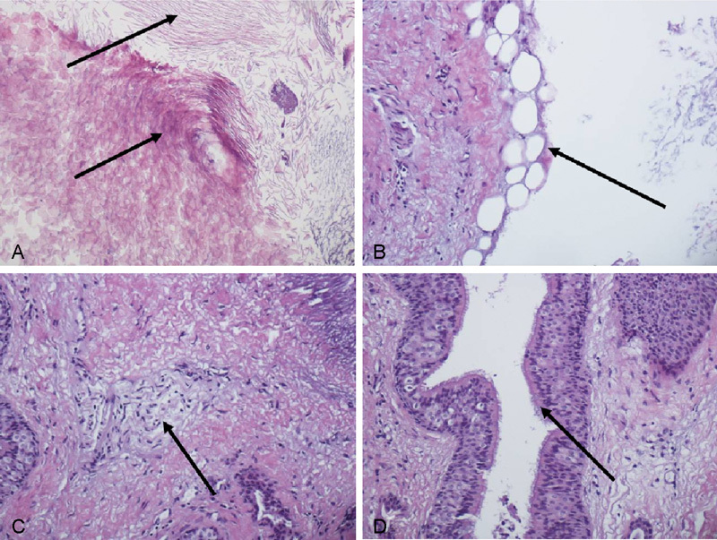
Microscopic images of the operative specimen demonstrating lamellar keratin debris with a foci of (A) anucleate squames, (B) adipose tissue, (C) peripheral nerve elements, and (D) respiratory-type epithelium with ciliated goblet cells.

Microscopic images of the operative specimen demonstrating lamellar keratin debris with a foci of (A) anucleate squames, (B) adipose tissue, (C) peripheral nerve elements, and (D) respiratory-type epithelium with ciliated goblet cells.