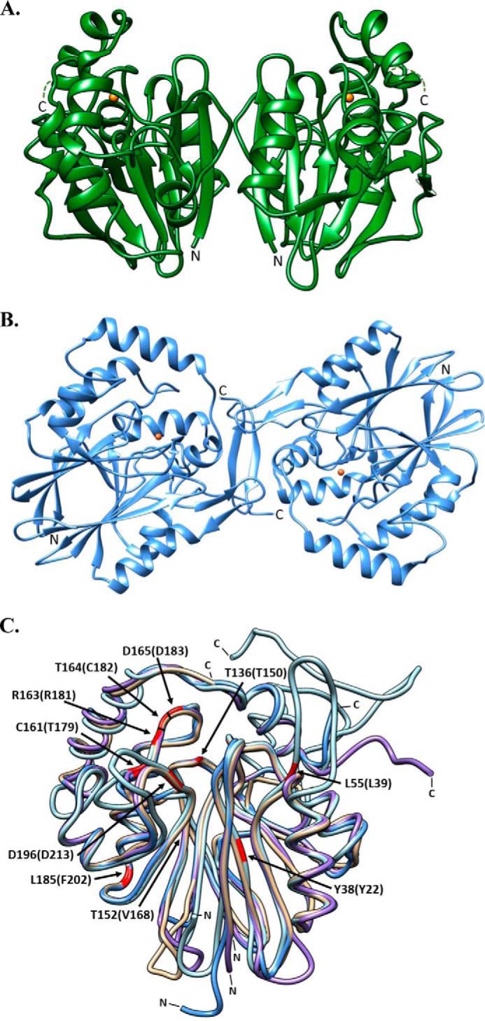
FIGURE 1.

Ribbon diagrams representing the oligomeric and superimposed structures of PDOs. A, PpPDO2 and B, MxPDO1b are represented in their dimeric states. The orange spheres in both enzymes represent Fe(II). C, the superimposed structures of MxPDO1b (tan), PpPDO2 (light blue), hPDO (blue), and AtPDO (purple). The corresponding N and C termini are marked N and C, respectively. Residues corresponding to ethylmalonic encephalopathy-causing mutations are highlighted in red. The residues are numbered according to the hPDO sequence, with equivalent residues from PpPDO2 in parenthesis. This figure was generated using Chimera (UCSF) (32).
