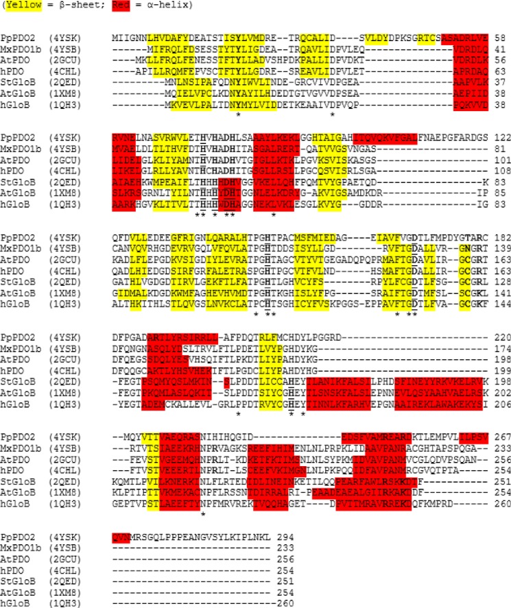
FIGURE 5.
Multiple sequence alignment of four PDOs and three glyoxalases II. Included in the alignment are PDOs from P. putida (PpPDO2), M. xanthus (MxPDO1b), human (hPDO), and Arabidopsis (AtPDO), as well as glyoxalase II enzymes from human (hGloB), A. thaliana (AtGloB), and Salmonella typhimurium (StGloB). The α-helices and β-strands are highlighted in red and yellow, respectively. Functionally significant residues are bolded, and residues involved in binding metals are underlined. Single conserved residues are marked with an asterisk, whereas conserved motifs are enclosed by a box. Multiple sequence alignment was performed with CLUSTALW2 using a BLOSUM weighting matrix. Secondary structure for each enzyme was calculated from PDB coordinates using DSSP (version 2.0)(29).

