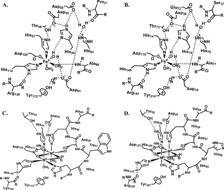
FIGURE 6.
Metal-binding and secondary coordination sphere architectures of PDOs and glyoxalases II. The two structures shown at the top panel correspond to PpPDO2 (A) and MxPDO1b (B). At the bottom panel are glyoxalase II enzymes from human (C) and S. typhimurium (D). The extensive hydrogen bond network that appears to be characteristic of PDOs is substituted with a conserved, one-turn α-helix (resides 56–60 of human and 55–59 of S. typhimurium) in the glyoxalases II that is involved in coordinating the second metal.

