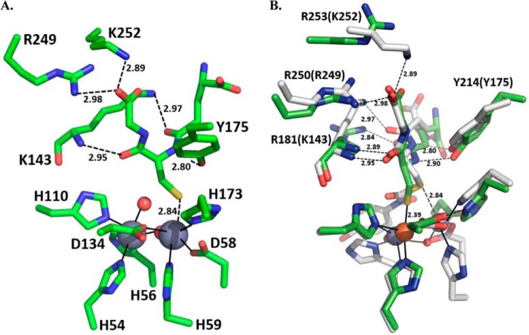
FIGURE 7.
Active site representations of PpPDO2 and human glyoxalase II in complex with GSH. A, the binding pocket of human glyoxalase II (Protein Data Bank code 1QH5) with a bound GSH molecule is shown. The two gray spheres represent the catalytic Zn(II) ions (28). B, for comparison, the GSH-binding modes and participating residues of PpPDO2 (green) and human glyoxalase II (gray) were superimposed. Residue labels that correspond to residues of the glyoxalase binding pocket are indicated in parentheses. Bond lengths are written near their respective bond representations. This figure was generated using PyMOL Molecular Graphics System, version 1.7.0.3 (Schrödinger, LLC).

