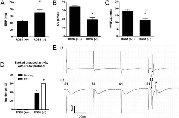
FIGURE 7.
The electrophysiology of isolated left atria. A, atrial effective refractory period. B and C, conduction velocity (B) and minimum wave front cycle length (C, mWFCL) were all slightly altered in left atrial tissue deficient in RGS4−/− mice compared with RGS4+/+ (n = 13 for RGS4+/+ and n = 8 for RGS4−/−; * p value <0.05 by t test). D, percentage of atria in which unpaced evoked activity was induced by S1-S2 protocol in absence or in presence of ET-1 in 13 RGS4+/+ and 8 RGS4−/− mice. * and #, p value <0.05 by Fisher's exact test versus RGS4+/+ mice in the absence and presence, respectively, of ET-1. E, representative traces of field potential recording during S1-S2 pacing protocol in RGS4+/+ (panel i) and RGS4−/− (panel ii) atria in the presence of 10 nm ET-1, with the latter showing evoked abnormal activity (*).

