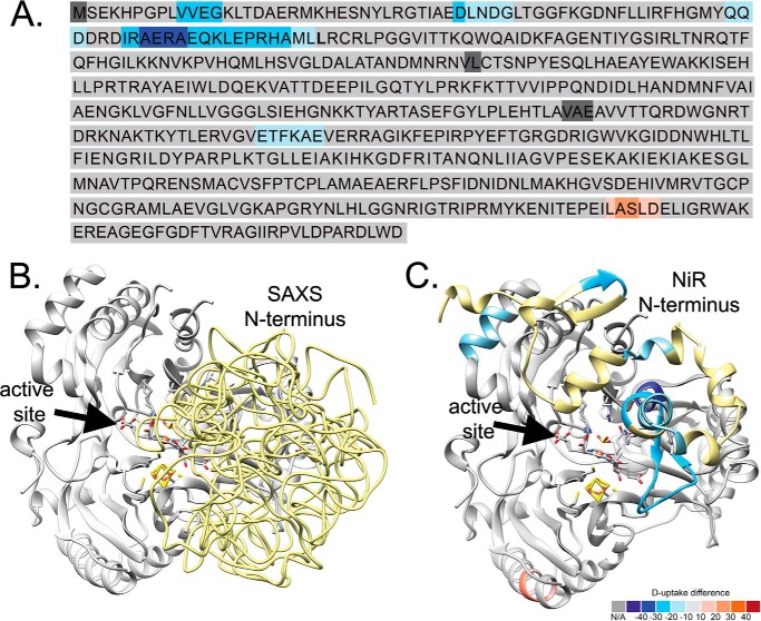
FIGURE 4.
Heat map for SiRHP HDX and SAXS modeling. HDX shows that the N terminus of SiRHP is buried in the SiR complex. A, sequence of SiRHP in which light-to-dark blue shows increasingly buried residues and pink-to-red shows increasingly exposed residues, measured by the absolute deuterium uptake. Leu-80 is in boldface type. B, six independent models of the N terminus in E. coli SiRHP built from a SAXS curve, superimposed, show that the compact N-terminal 80 amino acids (yellow, displayed as pseudochain models), which are missing in the x-ray crystallographic structure (9), sit over the active site, as in the NiR homolog (C). Colors are as in A, modeled from Protein Data Bank files 2AKJ (38) (yellow, N-terminal amino acids 1–80, as in B) and 2GEP (46) (gray, amino acids 80–570).

