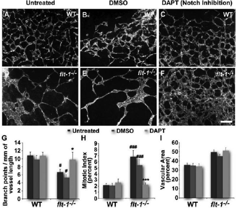
Figure 1. Notch inhibition by DAPT rescues the dysmorphogenesis of flt-1−/− blood vessels.
Wild-type (A-C) and flt-1−/− (D-F) day 8 ES cell-derived vessels stained for PECAM-1. Scale bar, 100 μm. Dy 8 vessel networks assessed for branch points per vessel length (G). #, p≤0.05 vs. WT of same treatment group. *, p≤0.05 vs. flt-1−/−/untreated or flt-1−/−/DMSO. Dy 7 vessel mitotic indicies were quantified by counting PH3+/PECAM-1+ cells and normalizing to total PECAM-1+ cells (H). ###, p≤0.0005 vs. WT of same treatment group. ***, p≤0.0005 vs. flt-1−/−/untreated or flt-1−/−/DMSO. Vessel area relative to total area for dy 8 ES cell-derived blood vessels (I). Values are averages +/− standard error of the mean (SEM).

