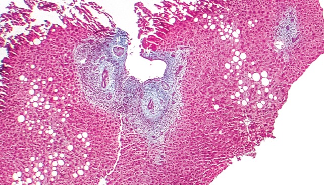
Fig. 3. Trichrome, X40; Liver Biopsy.
Low power view demonstrating a focal lesion typical for primary sclerosing cholangitis. Periductular layered fibrosis (characteristic “onion skin” pattern) is seen with edema and inflammation surrounding the interlobular bile ducts in the center of the field. A normal portal area with bile ducts is seen in the upper right corner of the image.

