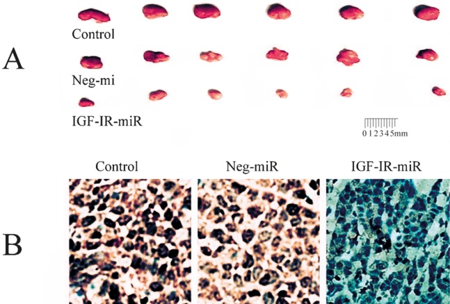
Fig. 2. Alterations of histopathology and immunohistochemistry in xenograft tumors.
(A) The size and gross features of xenograft tumors in nude mice from the different treatment groups: control group, PLC/PRF/5 cells transfected without any miR; the neg-miR group, PLC/PRF/5 cells transfected with neg-miR; the miR group, PLC/PRF/5 cells transfected with miR; (B) IGF-IR IGF-IR immunohistochemical analysis of the xenograft tumor tissues (SP, 400 ×) (unpublished data, Yao et al.).

