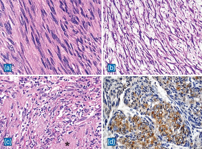
Fig. 3. Histpathological findings of IMT.
a: Cellular areas in fibromatosis-like pattern composed of elongated spindle cells with vesicular nuclei in a collagenous background (H&E, x400). b: Loosely arranged spindle cells in a myxoid background with scattered lymphocytes in myxoid-vascular pattern (H&E, x100). c: The compact spindle pattern with spindle cells arranged in a fascicular pattern in a collagenous stroma (asterisk). Numerous lymphocytes and plasma cells are present. (H&E, x200). d: Cytoplasmic staining of ALK in IMT of the liver (CD246, ALK protein counterstained with hematoxylin, x 200).

