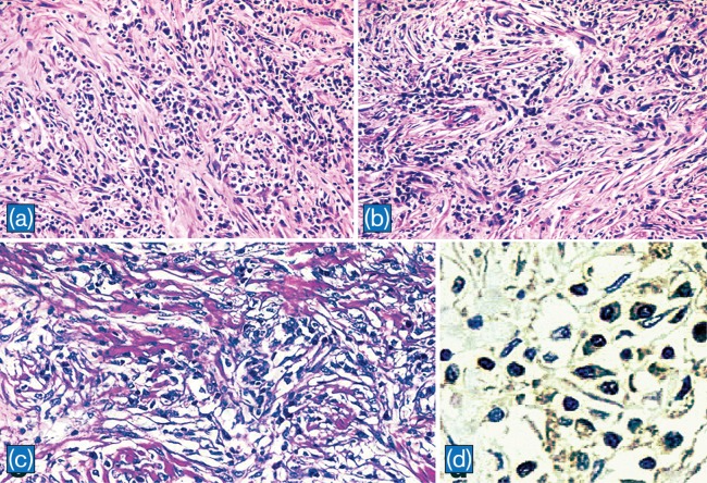
Fig. 4. Histpathological findings of IPT.
a: IPT composed of diffuse infiltration of lymphocytes and plasma cells in a background of collagen (H&E, x100). b: In some areas, lymphocytes and plasma cells are prominent around vascular structures (H&E, x100). c: In another case of IPT, spindle cells intermingled with lymphocytes, plasma cells, and histiocytes in a background of collagen deposits (x 200). d: IgG4 positive plasma cells in IPT presented in a and b (x 400)

