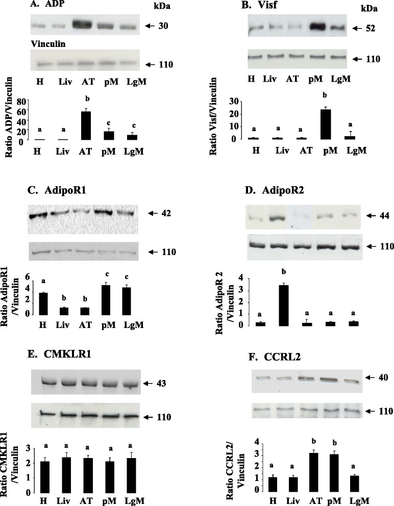
Fig. 2.

ADP (a), Visf (b), AdipoR1 (c), AdipoR2 (d), CMKLR1 (e) and CCRL2 (f) protein expression in peripheral tissues. ADP (a), Visf (b), AdipoR1 (c), AdipoR2 (d), CMKLR1 (e), and CCRL2 (f), protein levels were analyzed by western blotting in different peripheral tissues (Heart (H), Liver (Liv), Adipose Tissue (AT, pectoral (pM) and leg muscles (LgM) from six animals. Vinculin was used as a loading control. Data are represented as mean ± SEM (n = 6 animals). Different letters indicate a significant difference
