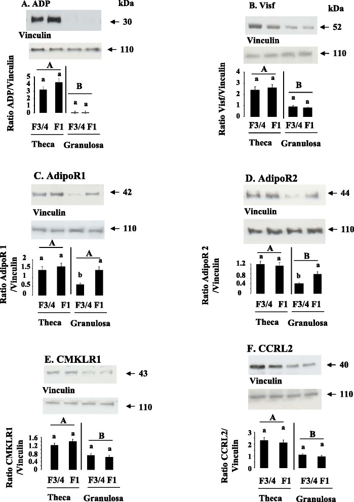
Fig. 5.

ADP (a), Visf (b), AdipoR1 (c), AdipoR2 (d), CMKLR1 (e), and CCRL2 (f), protein expression in ovarian cells. ADP (a), Visf (b), AdipoR1 (c), AdipoR2 (d), CMKLR1 (e), and CCRL2 (f), protein levels were analyzed by western blotting in theca and granulosa cells from F1 and F3/4 follicles. Vinculin was used as a loading control. Results are represented as means ± SEM (n = 6 animals). Different capital letters indicate a significant effect of the type of cells (Theca vs Granulosa cells) whereas lower case letters indicate a significant effect of the follicular stage (F3/4 vs F1). Data are represented as mean ± SEM. (n = 6 animals)
