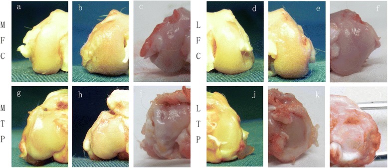
Fig. 3.

Typical macroscopic cartilage lesions among the groups in the four regions for the OA middle stage. The DHEA 9 w group exhibited a lower severity grade of macroscopic cartilage lesions in the 4 joint compartments compared with the NS 9 w group (P < 0.05). a-c represents the medial femoral condyles (MFC) of different groups (a: Sham, 9 w; b: ACLT+ DHEA, 9 w; c: ACLT + NS, 9 w). d-f represents the lateral femoral condyles (LFC) of different groups (d: Sham, 9 w; e: ACLT+ DHEA, 9 w; f: ACLT + NS, 9 w). g-i represents the medial tibial plateaus (MTP) of different groups (g: Sham, 9 w; h: ACLT+ DHEA, 9 w; i: ACLT + NS, 9 w). j-l represents the medial tibial plateaus (LTP) of different groups (j: Sham, 9 w; k: ACLT+ DHEA, 9 w; l: ACLT + NS, 9 w)
