
Fig. 1.
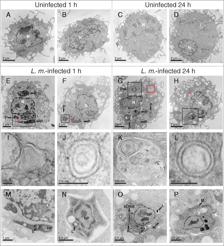
Ultrastructural investigation of autophagy induction in L. m.-infected BMDM with TEM. Methods: BMDM from BALB/c mice were infected with L. m. promastigotes for (e, f, i, j, m, n) 1 h and (g, h, k, l, o, p) 24 h. a–d Uninfected BMDM were incubated for the same amount of time in RPMI medium. All BMDM were subjected to TEM analyses. Results: Autophagic phenotypes characterized by (e–h) a strong vacuolization, (i, k) presence of MLS and (j, l) autophagosomes detected in L. m.-infected BMDM 1 h p.i. and 24 h p.i. compared to uninfected control BMDM. Details in images (i–p) were magnified from images (e–h) from sections of L. m.-infected BMDM (red squares = MLS in i and k, red circles = autophagosomes in j and l, black squares = intracellular parasites in m–p). K = kinetoplast, kDNA = kinetoplastid DNA, L = lysosome-like vacuole, M = mitochondrion, N = nucleus of macrophage, NP = nucleus of parasite, P = parasite