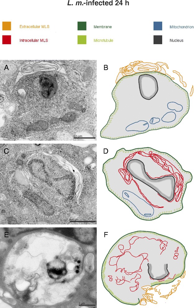
Fig. 5.

Ultrastructural investigation of parasite-associated localization of MLS with TEM. Methods: BMDM from BALB/c mice were infected with L. m. promastigotes for 24 h and subjected to TEM analyses. Results: MLS were frequently observed (a) in the cytoplasm of BMDM closely associated with intracellular amastigotes, and (c) localized in the cytoplasm of parasites. e Additionally, digested amastigotes with a defective cell membrane and remains of intracellular MLS were found in L. m.-infected BMDM 24 h p.i.. (b, d, f) Schematic illustrations of images (a, c, and e), respectively
