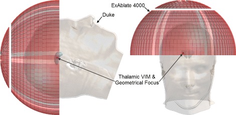
Fig. 2.

“Duke” anatomical head model and ExAblate®; 4000 transducer array model. The “Duke” anatomical head model and the ExAblate®; 4000 transducer array were used in this study. The positioning of the head model within the transducer as well as the location of the geometric focus in the right thalamic ventral intermediate (VIM) nucleus can be seen
