
Fig. 2.
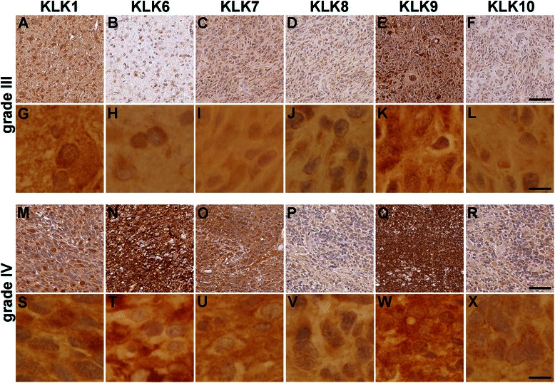
Immunohistochemical staining for KLK1, KLK6, KLK7, KLK8, KLK9 and KLK10 in astrocytomas. Representative photomicrographs of grade III astrocytomas (a–l) and grade IV astrocytomas (m–x) stained for KLK1 (a, g, m and s), KLK6 (b, h, n and t), KLK7 (c, i, o and u), KLK8 (d, j, p and v), KLK9 (e, k, q and w), or KLK10 (f, l, r and x). Images provided at low (a–f and m–r, scale bar = 50 μm) and high magnification (g–l and s–x, scale bar = 10 μm)