
Figure 2.
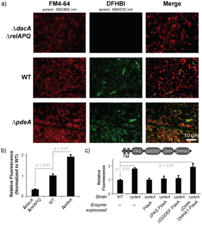
The yuaA-Spinach2 biosensor detects altered cyclic di-AMP levels in Listeria monocytogenes.(a) Fluorescence microscopy images of L. monocytogenes 10403s strains expressing yuaA-Spinach2 tRNAs after incubation with the membrane dye FM4-64 or DFHBI (left and middle panels, respectively; right panels show merge). (b) Flow cytometry analysis of L. monocytogenes strains expressing yuaA-Spinach2 tRNAs. Error bars represent standard deviation between four independent biological replicates. (c) Flow cytometry analysis of WT or ∆pdeA strains with no complementation or complemented with different pdeA enzyme constructs. The domain arrangement for PdeA is shown; the transmembrane (TM) domain was not included in the enzyme constructs. Error bars represent standard deviation between three independent biological replicates.