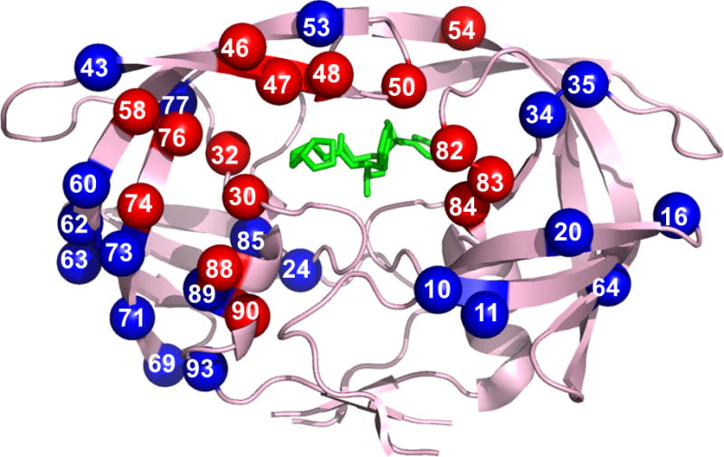
Figure 2. Protease dimer showing sites of resistance mutations.

Locations of resistance associated mutations mapped on the HIV protease dimer bound with DRV (PDB: 2IEN). The protease dimer is shown in pink ribbons with DRV in green sticks. Major and minor mutations are distributed among both monomers and shown in red and blue spheres, respectively.
Adapted with permission from [30] (2009); using updated resistance mutations from [21].
For color images please see online www.future-science.com/doi/full/10.4155/FMC.15.44
