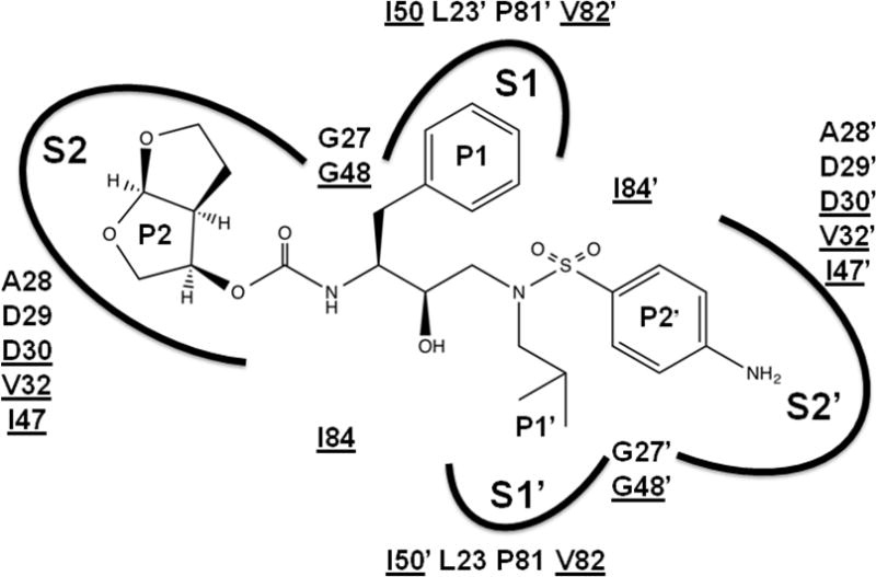
Figure 4. Darunavir in the binding site of HIV protease.

Darunavir is shown with groups labeled P2-P2′. The protease binding site is indicated by arcs for subsites S2, S1, S1′ and S2′ and the amino acid residues contributing to the binding site. Underlined amino acids are mutated in drug resistance.
