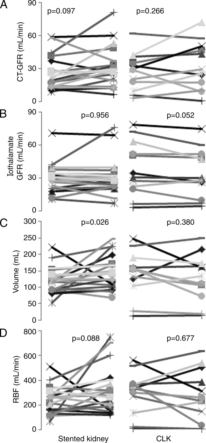
Figure 5:
Graphs of individual single-kidney GFR, volume, and RBF determined by means of CT or iothalamate clearance at baseline and at 3-month follow-up in the stented group. A, CT GFR, B, iothalamate GFR, C, kidney volume, and D, RBF are shown. Revascularization did not affect CT GFR, iothalamate GFR, or RBF (patients with stents, n = 16; kidneys with stents, n = 20; contralateral kidneys without stents [CLK], n = 12).

