
Figure 2a:
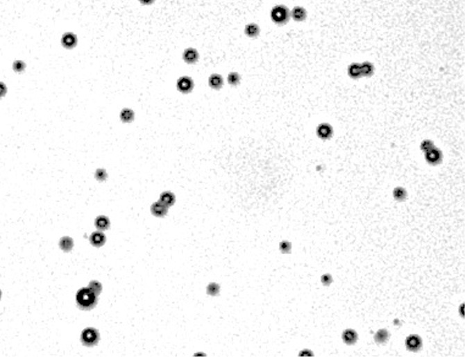
(a) Phase contrast and (b) fluorescence (fluorescein channel) microscopy images (original magnification, ×10) of MBs after DNA loading. SYBR Gold (Life Technologies), a dye that fluoresces in the presence of DNA, was added to both samples. Note the ring enhancement, indicating that DNA has been incorporated in the MB shell.