
Figure 3:
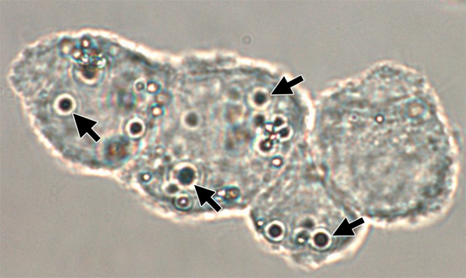
Confocal phase contrast microscopy image (original magnification, ×100) demonstrates C17.2 cells labeled with MBs (arrows). On average, each cell is associated with 2–4 MBs, which are either internalized or strongly adherent to the cell membrane.