Figure 1.

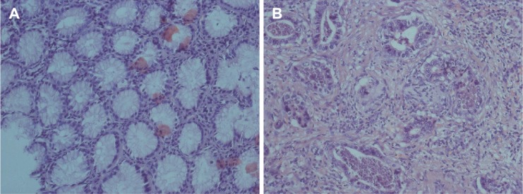
HE staining was performed using the sections of normal tissue and GC.
Notes: (A) The normal tissue specimens; (B) gastric adenocarcinoma (Hematoxylin-eosin staining, ×200).
Abbreviations: HE, Hematoxylin-eosin; GC, gastric cancer.

HE staining was performed using the sections of normal tissue and GC.
Notes: (A) The normal tissue specimens; (B) gastric adenocarcinoma (Hematoxylin-eosin staining, ×200).
Abbreviations: HE, Hematoxylin-eosin; GC, gastric cancer.