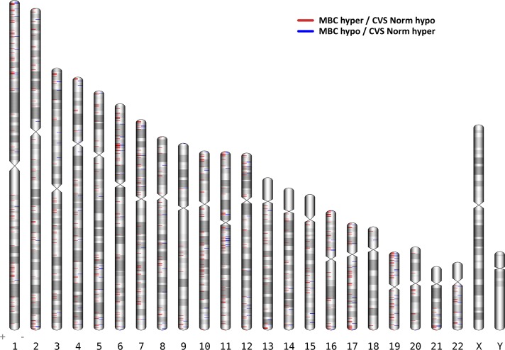
Fig 2. Ideogram of the 958 differentially methylated CpGs (DMCs) between Maternal blood cells (MBC) and Chorionic villus samples (CVS).
DMCs that are hypermethylated in MBCs and hypomethylated in CVS are shown in red., whereas DMCs hypomethylated in MBC and hypermethylated in CVS are blue. Chromosomes 13, 18 and 21 have been marked with a black triangle. DMCs on these chromosomes holds the possible use for diagnosis of the three main trisomies; Trisomy 13, 18 and 21.

