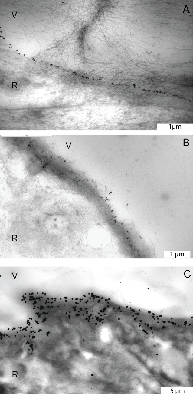
Fig 2. Transmission electron microscopic images of the pre-equatorial, equatorial and posterior ILM stained with an antibody against type IV collagen.
A. Pre-equatorial ILM of the eye of the 56 year-old donor. Note the linear distribution pattern of the labeling. B. Equatorial ILM of the eye of the 77 year-old donor. Note the more diffuse staining pattern. C. Posterior vitreoretinal interface of the 56 year-old donor eye. Type IV collagen appears to be distributed throughout the entire thickness of the ILM. A and B, bar = 1 μm; C, bar = 5 μm. V = vitreous; R = retina.

