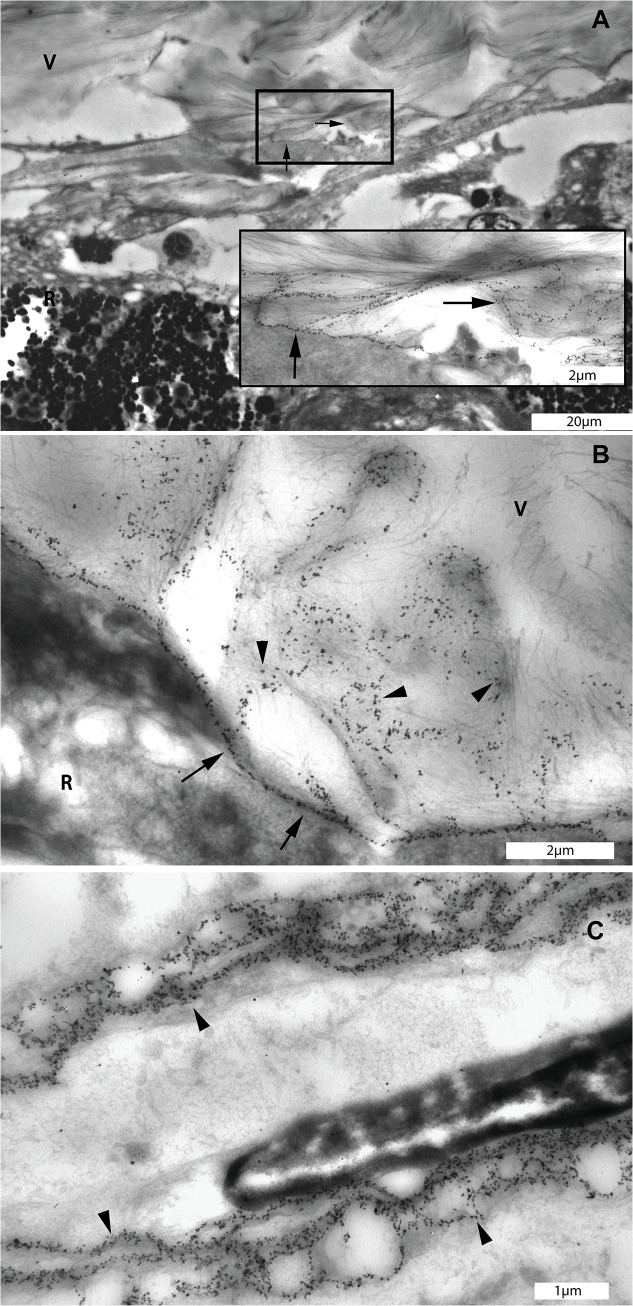
Fig 6. Transmission electron microscopic images of diffuse networks which were stained with type IV collagen antibody at the pre-equatorial vitreo-retinal interface of the eye of the 56 year-old donor.
A. Immunogold labeling of type IV collagen displayed a reticular pattern at the pre-equatorial vitreoretinal interface (square box). Insert: Detail of marked area in A: Type IV collagen positive structures in the vitreous extending themselves to the adjacent retina (black arrows). B: Linear distribution of type IV collagen indicating the location of the ILM (arrows) and type IV collagen positive structures in the vitreous attaching themselves to the ILM and superficial retina (black arrow heads). C. Distribution of type IV collagen in the basement membrane of a choroidal blood vessel displaying a similar reticular pattern as that found in the pre-equatorial intravitreal reticular structures (black arrow heads). A, bar = 20 μm, insert, bar = 2 μm; B, bar = 2 μm; C, bar = 1 μm. V = vitreous; R = retina.

